Dental diagnostics and treatment (oral cavity diagnostics, 3D dental diagnostics)Modern dentistry and cosmetology can correct many defects of the oral cavity, but first of all it is necessary to define patient’s state of health and treatment methods. For this purpose a dentist specialist should assess the state of teeth and gums. DENTIST&CO has all necessary tools and laboratory equipment to perform primary examination and accurate additional clinical and functional assessment. We value your health and time as well as our professional reputation. Therefore, we are trying to make the treatment plan as best and efficient as possible. To perform the required dental diagnostics and treatment we use different state of the art methodologies with application of the innovative equipment. * Computed tomography (CT) – «golden standard» of oral cavity diagnostics. Three dimensional x-ray image can provide a picture of the problem area and help to define optimum treatment program. KT is a safe method (ray load can be compared to the one during a long plane travel) and it is convenient tool for a patient, since it helps to assess the state and size of the bone, virtually “install” implants, model their location in the oral cavity and in the bone taking into consideration patient’s anatomic features. 3D-examination is indispensable during teeth treatment, since a dentist can get full information about the dental root system and possible bone inflammation, its size and location. * Panoramic imaging of teeth and temporomandibular joint - provides panoramic images containing useful information about the hard and soft tissues, position of teeth and bones and possible inflammation. 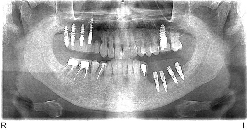 * Teleradiography (TRG) – helps to define peculiarities of the skull and facial bones, size and location of jaws, analyze growth of facial bones, identify problems and keep track of the dynamics of orthodontic treatment. 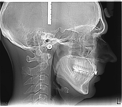 TRG provides much information to different specialists involved in the treatment process. It is indispensable to define individual’s bite, teeth abrasion and the requirement to restore optimum teeth height. It is one of the basic images used by the specialists at different stages of patient’s treatment. * Collecting information on patient’s complaints and anamnesis, filling out clinical and functional application is an important part of diagnostic examination, when the dentist specialist fills patient’s electronic card. It contains the information on allergy reactions, intolerance to some drugs as well as concomitant diseases. This information is needed to the specialists at different stages of dental reconstruction. If the patient experiences frequent headaches additional form should be filled, since this problem could be related to bite distortion and change of the distance between the jaws. * Diagnostic models – plaster dental/gum casts that allow the specialist to view the teeth position as well as tooth shape and angulation. * Analysis of jaws models in the articulator – hardware methodology that allows the dentist specialist to simulate multilateral movement of the lower jaw, visualize teeth contact, assess whether these movements and contacts are appropriate and decide whether correction is needed with the purpose to prevent further teeth abrasion. 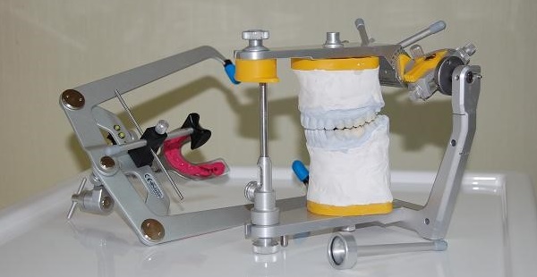 * Filling parodontologie card – the dentist specialist assesses the tissues around the teeth, their size, fixture to the bone and identifies possible gums bleeding. * JVA equipment allows the doctor to identify the pathological changes of the temporomandibular joint. 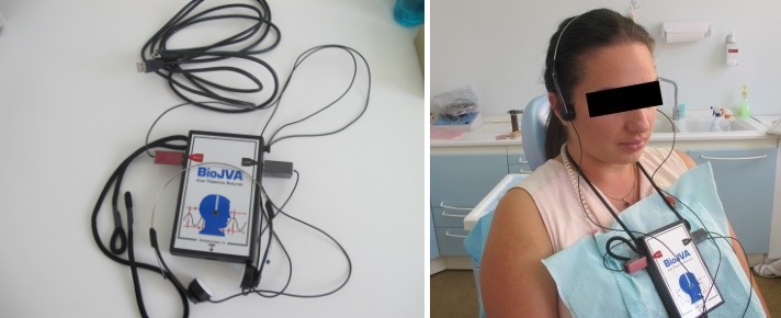 * Axiography – an effective method of oral cavity diagnostics used to record all mandibular movements (translations and rotations). 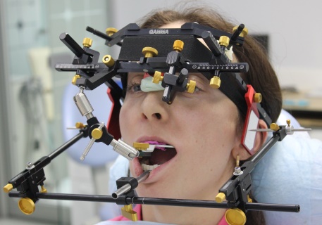 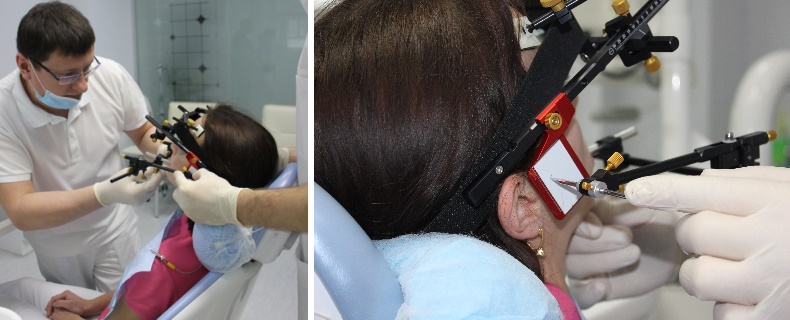 It provides the input information for the articulator (equipment simulating mandibular movement). Axiography is indispensable for restoration of the tooth row, since it allows the dental specialist to estimate with mathematical accuracy the position and tooth cusp angle of the future constructions. * Photo-statistical analysis – is performed with application of special dental cameras, flashlights and reflectors that allow the dental esthetic specialist to view the smallest details and transfer this information to the dental technician to ensure harmonic restoration of the whole system. This procedure is conducted at the beginning of the treatment process, it allows to analyze proportions of facial features as well as peculiarities of the hard and soft tissues. Photo-statistical analysis is conducted at all treatment stages. The information obtained is stored in patient’s card: it allows the specialists to control the dynamics of restoration of dentogingival and dentofacial esthetics. * T-SCAN – is an equipment used to assess the position and strength of occlusal contact at jaw movements, prevent crown and tooth tubercle fracturing as well as minimize adaptation to the constructions in the oral cavity, so that they could be “invisible” for a patient. 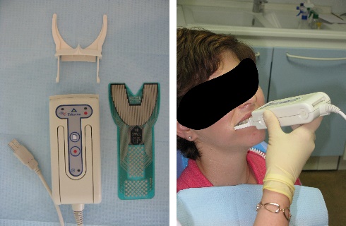 * WaxUp is used for provisional wax modeling of metal ceramic restorations that reflect the shape and size of the final restoration. You can specify all details and look at the restorations in the oral cavity. DENTIST&CO is also equipped with the state of the art equipment for 3D dental diagnostics: scanning the jaws, computer modeling, using 3D-printer to print temporary and permanent constructions. This significantly improves the accuracy - to a micron size. Please note! After developing the treatment plan, the dentist specialist agrees with the patient all required procedures and the treatment method. After this, the Dental Services Agreement shall be concluded. The patient receives the detailed information on the planned procedures and the their cost. Dental diagnostics and treatment are provided after the patient has signed the informed consent. Appointment with the patient for the future examination and additional control are scheduled on individual basis. |
|
Send your name and telephone number, we will ring up you and we will give free consultation